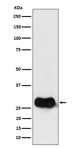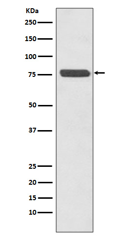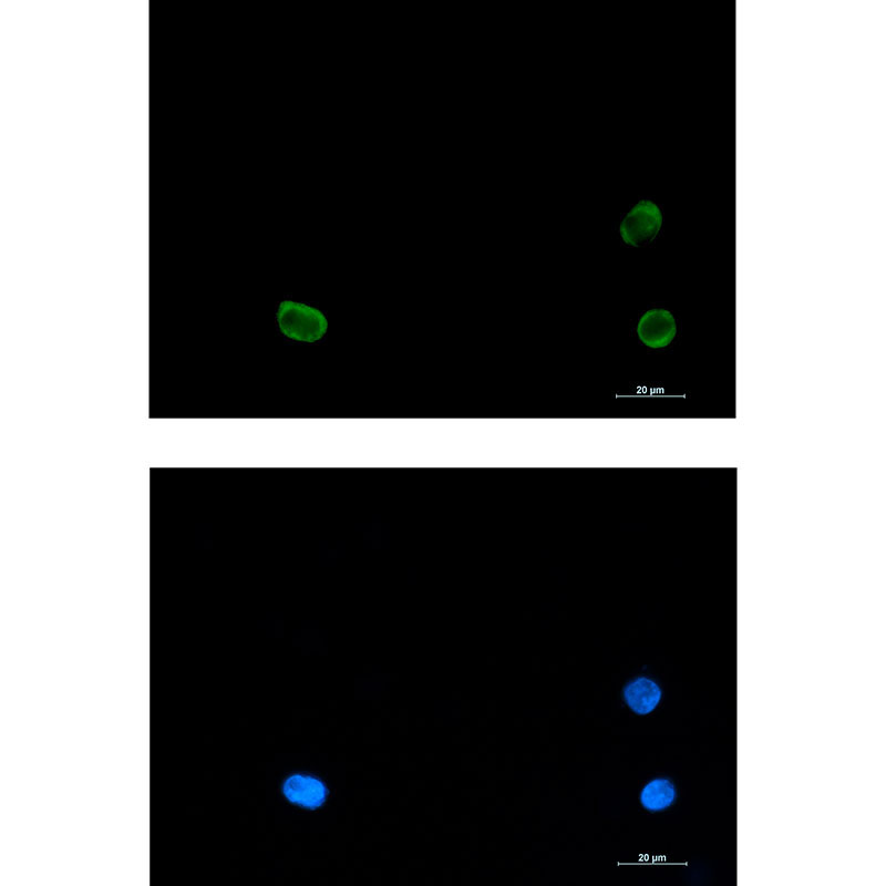
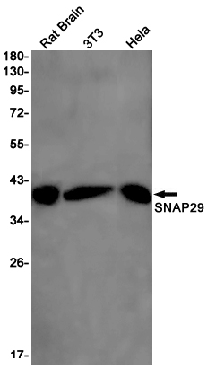
urrentScrollPos = maxScroll; } updateScrollPosition(); updateArrows(); }); // 左箭头点击 - 向左滚动 $('.img-arrow.left').click(function () { if ($(this).hasClass('disabled')) return; currentScrollPos -= SCROLL_DISTANCE; if (currentScrollPos < 0) { currentScrollPos = 0; } updateScrollPosition(); updateArrows(); }); // 初始化 setTimeout(function () { containerWidth = $(".scroll-wrap").width(); calcMaxScroll(); currentScrollPos = 0; updateScrollPosition(); updateArrows(); }, 100); // 窗口改变时重新计算 $(window).resize(function () { setTimeout(function () { containerWidth = $(".scroll-wrap").width(); calcMaxScroll(); if (currentScrollPos > maxScroll) { currentScrollPos = maxScroll; } updateScrollPosition(); updateArrows(); }, 200); }); });
SNAP29 Rabbit mAb

Item Number:R07108

Delivery Time:1-2 Days
One-click copy of product information
Contact Us
  • Product NameSNAP29 Rabbit mAb
  • synonymSNAP29
  • applicationWB;IP
  • reactive speciesHuman;Mouse;Rat
  • Molecular Weight29 kDa
  • Apparent Molecular Weight29 kDa
  • Immunogen/
  • Gene ID9342
  • Protein CodeO95721
  • host speciesRabbit
  • Antibody IsotypingIgG
  • purificationAffinity purification
  • storeStore at -20℃. Avoid freeze / thaw cycles.
  • Target BackgroundRelevant information can be queried against the UNIPROT database on the gene ID.
  • Recommended DilutionWB 1:1000-1:2000
    IP 1:20-1:50
  • Product formLiquid
  • Buffer SystemPBS with 1% BSA, 0.03% Proclin300 and 50% Glycerol.
🎯 1、Antibody selection and procurement
Q1 How to choose a primary antibody suitable for my experiment?

When selecting a primary antibody, the following factors should be comprehensively considered:

  • species reactivity:Confirm whether the antibody recognizes the species of your experimental sample (human, mouse, rat, rabbit, etc.)。
  • Application Type:Check the verified applications (WB, IHC, IF, IP, ChIP, Flow, etc.) listed in the antibody manual. Choosing verified applications can avoid the need for trial and error.
  • clone type:Monoclonal antibodies have strong specificity and small batch-to-batch variability; polyclonal antibodies have high sensitivity and recognize multiple epitopes, making them suitable for target proteins with low abundance.
  • Antigen Information:Confirm whether the immunogenic region recognized by the antibody includes specific structural domains, modification sites, or isoforms of the target protein.
Q2 What is the difference between monoclonal antibodies and polyclonal antibodies? How to choose?
Feature Monoclonal antibody Polyclonal antibody
Source Single B-cell clone Multiple B-cell clones
Specificity Very high, recognizing a single epitope Relatively high, capable of recognizing multiple epitopes
Sensitivity Medium Relatively high
Inter-assay consistency Excellent There are differences
Application preference IHC, flow cytometry, blocking experiment WB、IP、ChIP

Selection Recommendations: When high specificity is required (such as distinguishing between modification states) or long-term large-scale experiments are conducted, monoclonal antibodies are preferred; when detecting low-abundance proteins or exploring experimental conditions for the first time, polyclonal antibodies are more suitable.

Q3 How to interpret the "species reactivity" on the antibody instruction manual?

Species reactivity data is sourced from manufacturer validation or literature reports. If the instruction manual states "human, mouse", it indicates that the antibody has been validated to be positive in at least two species; if it only states "predicted", it means it is based solely on sequence homology analysis. When using across species, it is recommended to consult the literature or conduct validation on one's own.

Q4 I need to detect phosphorylated proteins. How can I select a phosphorylation-specific antibody?
  • Select antibodies that have undergone cross-validation for phosphorylated and non-phosphorylated proteins. The instructions should clearly demonstrate that the phosphorylated bands disappear after dephosphorylation treatment (such as lambda phosphatase).
  • Recombinant monoclonal antibodies are preferred due to their superior batch-to-batch stability.
  • Pay attention to whether the immune in situ points are consistent with the sequence of your experimental species.
❄️ II、Antibody storage and stability
Q5 How should antibodies be stored?
  • Undiluted antibody:According to the instructions, after aliquoting, the samples are generally stored at -20℃ (to avoid repeated freeze-thaw cycles), while some antibodies can be stored at 4℃ for short-term preservation. Glycerol protectant can enhance freeze-thaw stability.
  • Diluted antibody:It is recommended to prepare the working solution immediately before use. Working solutions should generally not be stored for extended periods. If storage is necessary, additives can be added to achieve a final concentration of 0.02% to 0.05% NaN₃ (not for use in live cell experiments) or commercial antibody stabilizer, can be stored at 4℃ for several weeks.
  • light-proof:Fluorescently labeled antibodies need to be stored in the dark, wrapped in aluminum foil or placed in a dark box.
Q6 What impact does repeated freeze-thawing have on antibodies? How can it be avoided?

Repeated freeze-thaw cycles can disrupt the spatial structure of antibodies, leading to aggregation, decreased activity, and even degradation. Ways to avoid this include:

  • Upon receipt of the antibody, it should be immediately aliquoted into small portions (e.g., 10 μL per tube), with each portion intended for single use only.
  • Label the packaging date and tube number, store at -20℃, thaw one tube before use, and it is not recommended to refreeze the remaining unused portion.
Q7 Can the antibody still be used if it precipitates or becomes turbid?
  • Slight precipitation:Centrifuge at 4℃ (10,000-14,000 g, 10 minutes), take a small aliquot of the supernatant to verify activity; or gently invert and mix (to avoid foaming).
  • A large amount of flocculent sediment:It may have denatured or aggregated, and continued use is not recommended.
  • Precautions:Some antibodies contain glycerol or BSA, which may appear slightly turbid, which is a normal phenomenon. Please refer to the instructions for further details.
🔬 III、Antibody usage and experimental optimization
Q8 What is the recommended dilution ratio for the antibody? How can it be optimized?

The recommended proportion in the instruction manual is only for initial reference. Optimization steps:

  1. Determine the initial dilution based on the type of application(WB:1:500-1:2000;IHC:1:100-1:500).
  2. Conduct a gradient dilution experiment(as 1:100、1:500、1:2000、1:8000)。
  3. Positive/negative controls are set simultaneously, and the optimal dilution is determined based on signal intensity and background signal-to-noise ratio.
  4. Adjust the incubation time (overnight incubation at 4°C can enhance the signal, while 1-2 hours at room temperature can reduce the background).
Q9 Why do multiple bands or non-specific bands appear in Western Blot?

Possible reasons and solutions:

  • The target protein exists in isoforms, undergoes degradation, or is modified:: Review the literature to confirm the expected band position and detect whether the protease inhibitor has lost its efficacy.
  • Excessively high antibody concentration:Increase the dilution ratio.
  • Insufficient specificity of primary antibody:Replace the antibody or use blocking peptide for pre-adsorption verification.
  • Secondary antibody cross-reactivity:Use a more specific secondary antibody (such as one that only binds to light chains), or increase the number of washing steps.
  • Excessive protein loading:Reduce the sample size.
Q10 How to deal with excessive background staining in IHC/IF experiments?
Reason Solution
Insufficient sealing Replace the blocking solution (5% normal serum + 1% BSA + 0.3% Triton) and extend the blocking time (1 hour at room temperature or overnight at 4°C)
Primary antibody concentration is too high Increase dilution ratio
Secondary antibody non-specific binding Set up no primary or secondary antibody control; use isotype control or pre-adsorbed secondary antibody
Endogenous peroxidase (IHC-HRP) Add an H₂O₂ blocking step (0.3% H₂O₂ for 10-15 minutes)
Intrinsic fluorescence (IF) Use a spontaneous fluorescence quencher (such as TrueBlack)
Q11 What are the special requirements for selecting antibodies in IP experiments?
  • The primary antibody must be able to recognize the natural conformational epitope, and cannot be selected solely based on WB verification results.
  • It is recommended to use antibodies coupled to protein A/G agarose or magnetic beads (direct method), or to incubate with the antibody first and then add the beads (indirect method).
  • Set up an isotype IgG control to exclude non-specific binding.
  • Ensure that the elution conditions do not damage the target protein (such as neutralizing immediately after elution with low pH glycine).
Q12How to choose antibodies in flow cytometry (Flow)?
  • Choose antibodies that have been validated for flow cytometry, and pay attention to the notation "Flow" or "FACS" in the instruction manual.
  • Surface staining uses live cells; intracellular staining requires fixation and membrane disruption.
  • When using fluorescently labeled secondary antibodies, pay attention to the compensation for channel overlap.
  • Set up the following controls: unstained control, isotype control, and single-dye compensation control (for multicolor experiments).
IV、Antibody specificity and verification
Q13How to verify the specificity of an antibody?

Cross-validation using multiple methods:

  • Knockout/knockdown validation:Using CRISPR/Cas9 to knock out cells or siRNA to knock down samples, the antibody signal should disappear or significantly weaken.
  • Multi-species orthologous protein detection:The antibody should recognize the target band of the expected size.
  • Peptide competition experiment:Pre-incubating immunogenic peptides can block specific signals.
  • orthogonal method:Compare the results with those obtained from mass spectrometry, ELISA, or another independent antibody.
Q14Why does the performance of the same antibody vary across different batches?

Inter-assay variability is a common issue in the production process, especially in polyclonal antibodies. Methods to reduce variability include:

  • Recombinant monoclonal antibodies (almost consistent between batches) are preferred.
  • Request the same batch of retained samples or reserve a sufficient amount from the supplier.
  • After receiving the new batch of antibodies, first perform parallel verification with the standard and the old batch.
Q15 Can I try using application types that are not listed in the manual?

Yes, but there are risks involved. Many antibodies may be effective in non-validated applications, but they require self-optimization and rigorous control settings. If successfully used, it is recommended to indicate in the publication that "this antibody XX application is validated by the laboratory itself".

🔧 V、Troubleshooting of common faults
Q16 What should I do if there is no signal or the signal is too weak in WB?

Step-by-step troubleshooting:

  1. Has the protein been successfully transferred to the membrane? Check with ponceau staining.
  2. Is the primary antibody ineffective? Try using a positive control lysis buffer for testing.
  3. Is the dilution too high? Reduce the dilution ratio and try again.
  4. Is the antigen epitope blocked? Consider changing the blocking buffer (e.g., using a protein-free blocking buffer or a different brand of skim milk powder).
  5. Insufficient detection sensitivity? Switch to a high-sensitivity ECL substrate or use a fluorescent secondary antibody.
Q17 The positive control is very strong, but there is no signal in the experimental group. What could be the reason?
  • The expression level of the target protein in the sample is extremely low: enrich the sample (such as running WB after IP) or use a more sensitive detection method.
  • Protein degradation: Prepare fresh lysis buffer and add protease/phosphatase inhibitors.
  • Species mismatch: Confirm the species homology between the antibody and the experimental sample.
Q18 What should I do if the IHC staining results do not match the expected localization?
  • Examine antigen retrieval methods (thermal retrieval and enzymatic retrieval have different effects on different antigens).
  • Verify whether the antibody recognizes the correct cellular structure in IHC (by consulting published literature or databases such as Human Protein Atlas).
  • Ensure that the tissue is fixed adequately but not excessively (10% neutral formalin, 24-48 hours).
📚 VI. Other common issues
Q19 How to inquire about the citation status of antibody-related literature?
  • The "References" section on the supplier's official website.
  • CiteAb(citeab.com)and Antibodypedia Provide cross-brand antibody citation rankings.
  • Google Scholar or PubMed Search directly for the antibody name or product number.
Q20 Can antibodies be used across species (such as using anti-human antibodies in mice)?

Two conditions must be met: ① the target protein exhibits high sequence homology between mice and humans; ② the instruction manual does not indicate "human specificity". Even so, it is still recommended to set up a positive control (such as a sample known to express the protein in mouse cell lysate) for verification.

Q21 What special properties are required for antibodies used in ChIP?

ChIP requires antibodies to recognize the natural chromatin epitopes after fixation. Selection criteria:

  • The instructions have clearly validated the application of ChIP or ChIP-seq.
  • Provide ChIP-qPCR positive primers or positive control antibodies (such as anti-H3K4me3).
  • Monoclonal antibodies are preferred to reduce non-specific enrichment.
Q22 How do I choose a fluorescent secondary antibody to match my microscope or flow cytometer?
  • Check the excitation light source and filter configuration in the instrument manual.
  • Common combinations include: DAPI (Ex 358 nm/Em 461 nm), FITC (Ex 488/Em 520), Cy3 (Ex 550/Em 570), and Alexa Fluor 647 (Ex 650/Em 670).
  • For multicolor experiments, it is necessary to select fluorescent dyes with spectral separation and prepare a single-dye compensation control.
🤝 VII、After-sales Service and Technical Support
Q23 What if the antibody doesn't work?

First, conduct a self-check according to the aforementioned troubleshooting guide. If the issue persists, contact the manufacturer's technical support and prepare the following information:

  • Antibody product number, batch number, and storage records.
  • Detailed experimental steps (blocking solution, dilution, incubation conditions, washing times, etc.)
  • Images of the results for positive and negative controls.

Most manufacturers offer quality guarantees, and upon verification, they may provide free replacements or refunds.

Q24 How can I obtain technical support and the latest validation data?
  • Visit the product page on the manufacturer's official website to download the latest version of the instruction manual and COA.
  • Contact technical support via email or phone, as some manufacturers offer WeChat/online customer service.
  • Subscribe to product update notifications to receive notifications of new validated applications and literature citations.